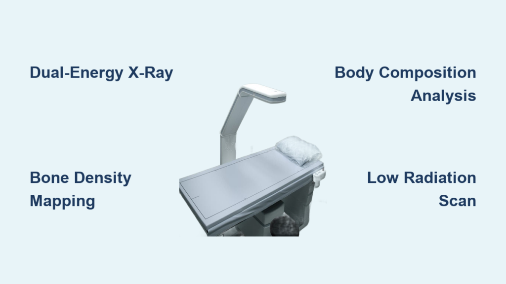
If you’ve ever wondered how doctors detect early bone loss or athletes precisely track body fat, the answer often lies in a DEXA scan. Dual-energy X-ray absorptiometry (DEXA or DXA) is the gold standard for measuring bone mineral density (BMD) and body composition, offering unmatched accuracy in clinical and research settings. Unlike standard X-rays that only reveal bone damage after significant loss, DEXA detects subtle changes long before fractures occur, making it essential for preventing osteoporosis and managing metabolic health.
But how does a DEXA scanner work? At its core, the technology uses two low-dose X-ray beams at different energy levels to distinguish between bone, fat, and lean tissue. By analyzing how these beams pass through the body, the scanner creates detailed maps of your skeleton and soft tissues, delivering precise measurements in under 20 minutes. Whether you’re being evaluated for fracture risk or optimizing athletic performance, understanding how DEXA works can help you appreciate its role in proactive health management.
The Dual-Energy X-Ray Principle
DEXA relies on the differential absorption of X-rays by tissues at two distinct energy levels. This dual-energy method allows the scanner to separate bone from soft tissue and further classify fat versus lean mass.
Why Two X-Ray Beams Each Tissue Absorbs X-Rays Differently
Each tissue type absorbs X-rays differently based on its atomic composition. Bone, which is high in calcium, absorbs more X-rays. Fat, being low density and carbon-rich, absorbs less. Lean tissue, which is water and protein-based, falls in between these two extremes.
Using just one X-ray energy would not allow accurate separation of these components. However, with two energies, the system solves two equations simultaneously to isolate each tissue type with remarkable precision.
The Mathematical Science Behind Tissue Separation
The fundamental physics follows the exponential attenuation law, where transmitted X-ray intensity equals the initial intensity multiplied by e raised to the power of negative mu times M. The variable I represents transmitted X-ray intensity, I0 represents initial intensity, mu represents the mass attenuation coefficient which varies by tissue and energy, and M represents areal density measured in grams per square centimeter.
With low-energy beams around 40 keV and high-energy beams around 70 keV, the scanner collects two data points per pixel. These are used to solve for bone mineral content and soft tissue density. Once bone is quantified and subtracted, the remaining soft tissue signal is analyzed using known fat-to-lean attenuation ratios, enabling a three-compartment model of the body.
This mathematical precision is what makes DEXA far more accurate than skinfold tests or bioelectrical impedance analysis for body composition measurement.
Images Generated During a DEXA Scan

A single DEXA scan produces multiple co-registered images, each revealing different aspects of your physiology. These images are aligned pixel-for-pixel, allowing direct comparison across tissue types from one scan session.
X-Ray Attenuation Image
The X-ray attenuation image shows raw transmission of X-rays through the body. It appears as a grayscale silhouette where areas appear darker where more radiation was absorbed, such as in bone, and lighter in soft tissue areas. This image is not used for diagnosis but helps technicians verify proper positioning.
Bone Mineral Density Map
The BMD map highlights only skeletal regions. It is color-coded or displayed in grayscale based on bone density values measured in g/cm². Clinicians use this to spot low-density zones, especially in the spine and hip, which are key sites for fracture risk assessment.
Body Composition Color Map
The body composition color map displays fat in red or yellow shades, lean mass in blue, and bone in white. This enables regional analysis comparing left versus right limb, measuring visceral fat, and calculating android to gynoid ratios. This capability is essential for tracking muscle symmetry in athletes or monitoring fat redistribution in metabolic disorders.
Key Metrics Reported by DEXA
DEXA does not just show pictures. It delivers quantitative data crucial for diagnosis and monitoring treatment progress over time.
| Measurement | What It Tells You | Unit |
|---|---|---|
| BMD | Bone strength per area | g/cm² |
| BMC | Total mineral in region | grams |
| Bone Area | Surface area scanned | cm² |
| Fat Mass | Total adipose tissue | kg or g |
| Lean Mass | Muscle, organs, fluids | kg or g |
| % Body Fat | Fat relative to total weight | % |
These values can be reported for the whole body, the spine from L1 to L4, the hip including the femoral neck and total hip, and individual arms, legs, or trunk regions. For example, a decline in femoral neck BMD over time signals rising hip fracture risk even if no symptoms exist.
Step-by-Step Scanning Procedure
Getting a DEXA scan is quick, painless, and requires minimal preparation. Understanding what happens during the scan helps patients feel more comfortable.
Patient Preparation Tips
Patients should wear loose, metal-free clothing such as clothing without zippers, belts, or underwire bras. All jewelry, phones, and keys should be removed before the scan. No fasting or injections are needed. Patients should inform staff if they are pregnant or have had recent contrast scans involving barium or CT with iodine, as scans are typically delayed 7 to 10 days after contrast procedures to avoid interference.
What Happens During the Scan
The patient lies flat on a padded table. A C-arm scanner moves slowly above the patient, emitting X-rays from below. The patient must stay completely still because even small movements blur results. The arm passes over the body in 10 to 20 minutes depending on the scan type. Most patients describe the experience as similar to getting a standard X-ray but easier.
Common Scan Sites and Their Purpose
Different scan sites serve different clinical purposes. The lumbar spine detects trabecular bone loss early. The hip femoral neck provides the best predictor of hip fracture risk. The forearm is used when the spine or hip cannot be scanned due to arthritis or implants. Whole body scans measure body composition for obesity evaluation, sports performance, and eating disorder assessment.
Peripheral DEXA devices that scan the wrist or heel are available but are less accurate than central machines.
Inside the Machine: DEXA Hardware Components
Every DEXA scanner combines precision engineering with advanced software to deliver accurate results. Understanding the hardware helps explain why DEXA is so reliable.
X-Ray Generator
The X-ray generator produces dual-energy beams via a tungsten-target X-ray tube. Voltage switching or filtration creates low and high energy pulses. Modern systems use pulsed beams that alternate energies rapidly during the scan for better accuracy.
Beam Collimation System
The beam collimation system shapes the beam into a fan or pencil pattern. This reduces scatter radiation and improves resolution. The system must align perfectly with the detector to maintain measurement accuracy across all scan types.
Detector Array
The detector array is located opposite the X-ray source. It captures transmitted photons and converts them into electrical signals. The system outputs a grayscale value per pixel where darker values indicate more absorption, such as in bone, and lighter values indicate less absorption, such as in fat.
Scanning C-Arm and Bed
The scanning C-arm holds the X-ray source and detector in precise alignment. It moves smoothly along rails above the patient to ensure consistent geometry across scans for reproducibility. The scanning bed features an open, flat design that reduces claustrophobia, supports patients up to approximately 450 pounds depending on the model, and provides easy access for elderly or mobility-impaired individuals.
Console and Analysis Software
The console controls scan acquisition and processing. The software runs algorithms to separate bone from soft tissue, trace bone edges automatically, calculate BMD, fat percentage, and lean mass, and flag poor-quality scans for retake. Advanced systems include automated rejection features if motion or artifacts compromise data quality.
Types of DEXA Scanners and Their Uses
Not all DEXA machines serve the same purposes. Different models are designed for different clinical needs.
Central DEXA
Central DEXA units are found in hospital radiology departments and bone clinics. They scan the hip, spine, and whole body with the highest precision available. These systems are the gold standard for diagnosing osteoporosis and monitoring treatment response. These large systems cost over $100,000 and require trained technologists to operate.
Peripheral DEXA
Peripheral DEXA devices are portable and compact, weighing approximately 60 pounds. They scan the wrist, heel, or finger and are commonly used for screening in pharmacies and community health fairs. The limitation is that peripheral devices cannot diagnose osteoporosis definitively and only suggest risk. Abnormal results require confirmation with central DEXA scanning.
Preclinical DEXA
Preclinical DEXA units are designed for animal research with mice and rats. They use ultra-low doses and provide rapid scanning in approximately 25 seconds. These units feature fully shielded cabinets and are used extensively in drug development and aging studies.
While quantitative ultrasound is also used for screening, it lacks DEXA’s accuracy and cannot measure body composition at all.
Interpreting Results: T-Score vs Z-Score
Your DEXA report includes standardized scores that compare your bone density to reference populations. Understanding these scores is essential for interpreting your results correctly.
T-Score
The T-score compares your BMD to healthy young adults at peak bone mass. It is used for men and women over 50. The World Health Organization defines T-scores as follows. A T-score of negative 1.0 or higher is normal. A T-score between negative 1.0 and negative 2.5 indicates osteopenia, which means low bone mass. A T-score of negative 2.5 or lower indicates osteoporosis. A T-score of negative 2.5 or lower combined with a fragility fracture indicates severe osteoporosis.
A T-score of negative 2.0 means your bones are two standard deviations below peak young adult density, increasing fracture risk up to four times.
Z-Score
The Z-score compares your BMD to people of your same age, sex, and ethnicity. It is used for children, adults under 30, and evaluating unusual bone loss. A Z-score below negative 2.0 suggests secondary causes such as malnutrition, hormonal disorders, or medication effects.
A normal T-score does not guarantee no fracture risk exists. Clinical tools like FRAX combine BMD with age, weight, smoking status, and prior fractures to estimate 10-year hip fracture probability.
Clinical Applications Beyond Osteoporosis
DEXA’s uses extend well beyond diagnosing brittle bones into many areas of medicine and wellness.
Primary Medical Uses
DEXA is used for osteoporosis diagnosis and monitoring, fracture risk prediction for hip and spine, tracking treatment response to bisphosphonates and hormone therapy, and assessing body composition in chronic diseases. These diseases include obesity, metabolic syndrome, sarcopenia which is age-related muscle loss, cancer cachexia, and HIV-associated fat redistribution.
Specialized Medical Indications
DEXA is particularly important for patients with hyperparathyroidism, where forearm scans are preferred because cortical bone is affected first. It is also critical for patients on long-term steroid use exceeding three months, as they face high risk of rapid bone loss. Patients with early menopause or oophorectomy without hormone replacement therapy experience accelerated BMD decline. People with rheumatoid arthritis, celiac disease, and thyroid disorders face secondary osteoporosis risks. Anyone with a prior fragility fracture has a major red flag for future fractures.
Athletes and fitness professionals also use DEXA to track muscle gain and fat loss, assess symmetry such as detecting limb imbalances, and optimize training and nutrition plans.
Radiation Safety and Limitations
Despite using X-rays, DEXA is extremely safe and well-tolerated by most patients.
Radiation Exposure Level
DEXA delivers 0.001 to 0.03 mSv per scan, which is equivalent to 1 to 3 days of natural background radiation. This is less than 1% of a chest X-ray. The low dose makes DEXA safe for repeat scans every 1 to 2 years to monitor change over time. No special shielding is typically required, and lead aprons are avoided because they interfere with imaging.
When to Avoid DEXA
Pregnancy is not a strict contraindication due to the low dose, but scans are postponed unless urgent. Recent contrast studies including barium or CT with iodine can mimic high bone density, so patients should wait 7 to 10 days after contrast procedures before scanning.
Known Limitations
DEXA cannot differentiate osteoporosis from osteomalacia, which is soft bones due to vitamin D deficiency. Spinal degeneration, arthritis, or prior fractures may falsely elevate BMD readings. Very large patients may exceed table weight limits or cause image distortion. Positioning errors affect reproducibility, which is critical for tracking change over time. Machine calibration is vital, and follow-up scans should ideally be performed on the same device.
Despite these limitations, DEXA remains the most validated and reproducible method for BMD and body composition measurement.
Evolution of DEXA Technology
DEXA did not emerge overnight. It evolved from earlier bone measurement techniques through decades of innovation.
The Birth of Dual-Energy Imaging
In the 1970s, DEXA technology was pioneered by Dr. Richard Mazess and Dr. Richard Cameron. They overcame limitations of single-energy absorptiometry, which could not separate bone from soft tissue. They introduced dual-photon absorptiometry first, then upgraded to X-rays for better speed and resolution.
Key Innovations Over Time
Two-energy X-ray pulses enabled pixel-level tissue differentiation. Improved edge detection allowed accurate bone boundary tracing. Faster scanning and lower radiation improved patient experience and safety.
Modern Advances
Fan-beam technology replaced pencil beams for faster whole-body scans. Enhanced software now includes AI-assisted segmentation and vertebral fracture assessment. Applications have expanded into sports science, nutrition, and aging research.
Advantages That Make DEXA the Gold Standard
DEXA remains the preferred method for bone density and body composition measurement due to its unique combination of accuracy, safety, and practicality.
Proven Benefits
DEXA offers the highest precision, detecting changes as small as 1 to 2% over time. It delivers the lowest radiation compared to other imaging methods. The procedure is fast and non-invasive with no needles or discomfort. It provides objective data that replaces guesswork with exact numbers. It tracks change effectively, making it ideal for monitoring treatment response. It delivers comprehensive output measuring bone, fat, and muscle in one scan. It is validated across diverse populations including men, women, the elderly, and athletes.
Comparison With Other Methods
Compared to bioelectrical impedance, which is highly variable and affected by hydration, DEXA provides consistent results. Compared to skinfold calipers, which are operator-dependent and inaccurate in obesity, DEXA offers precision. Compared to CT and MRI, which are accurate but expensive with high radiation from CT and less accessibility, DEXA strikes the ideal balance of accuracy, safety, speed, and cost-effectiveness.
Frequently Asked Questions About DEXA Scanners
Is a DEXA scan safe?
Yes, DEXA scans are extremely safe. The radiation dose is equivalent to only 1 to 3 days of natural background radiation, which is less than 1% of a standard chest X-ray. This makes it safe for repeated scans to monitor changes over time.
How long does a DEXA scan take?
A DEXA scan typically takes 10 to 20 minutes depending on the type of scan being performed. Whole-body composition scans may take longer, while focused hip or spine scans are usually faster.
Does a DEXA scan hurt?
No, a DEXA scan is completely painless. You simply lie flat on a table while the scanning arm moves over your body. There is no contact, no needles, and no discomfort involved.
What should I wear to a DEXA scan?
Wear loose, metal-free clothing without zippers, belts, buttons, or underwire bras. Remove all jewelry, watches, and phones before the scan. Some facilities provide a hospital gown if needed.
Can DEXA measure body fat percentage?
Yes, DEXA provides precise measurements of body composition including total fat mass, lean mass, and body fat percentage. It is considered more accurate than skinfold tests or bioelectrical impedance analysis.
How often should I get a DEXA scan?
For bone health monitoring, doctors typically recommend DEXA scans every 1 to 2 years if you are being treated for osteoporosis or osteopenia. For athletes tracking body composition, frequency varies based on goals and training programs.
Key Takeaways for Understanding DEXA Scanners
DEXA is more than a diagnostic tool. It is a window into your body’s structural and metabolic health. By harnessing the physics of dual-energy X-rays, it delivers precise, actionable insights that empower early intervention, personalized treatment, and long-term wellness.
The technology uses two low-energy X-ray beams to distinguish between bone, fat, and lean tissue with remarkable accuracy. This enables three-compartment analysis that no other simple method can match. Whether you are managing bone disease, optimizing athletic performance, or studying human physiology, understanding how a DEXA scanner works reveals why it remains the gold standard in body composition and bone density assessment.