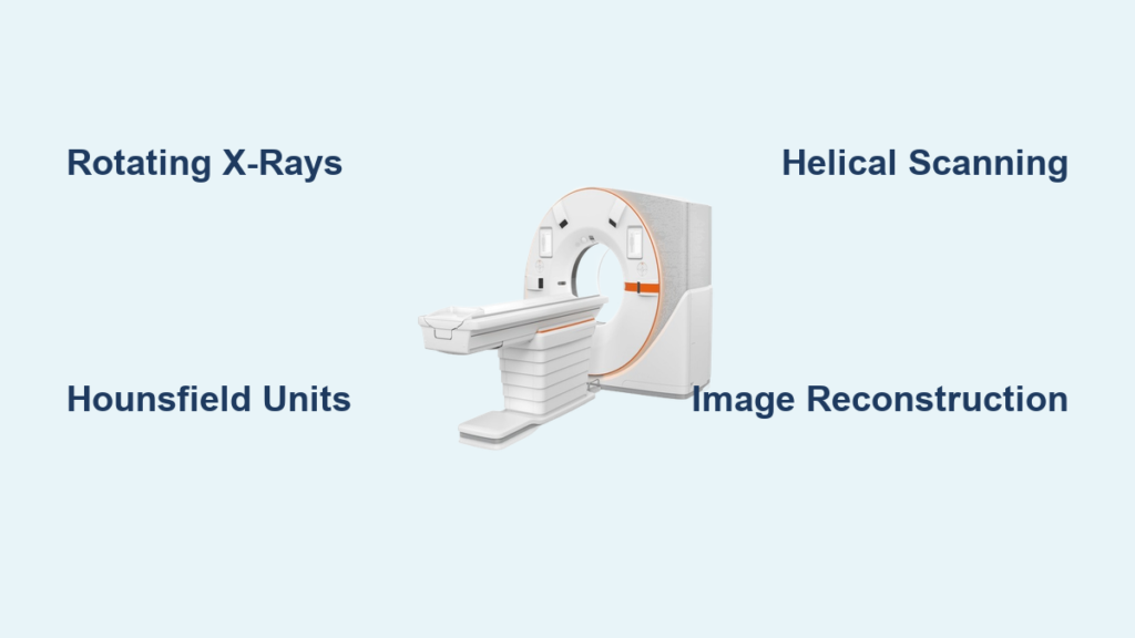
If you’ve ever wondered how doctors see inside the body without making a single incision, a CAT scanner—more accurately known as a CT (Computed Tomography) scanner—is one of the most powerful tools that makes it possible. This advanced imaging technology creates detailed cross-sectional views of bones, organs, blood vessels, and soft tissues, helping diagnose everything from broken bones to life-threatening cancers. But how does a cat scanner work? At its core, it combines rotating X-rays, high-speed detectors, and sophisticated computer processing to turn invisible radiation into life-saving images.
Unlike traditional X-rays that produce flat, 2D images with overlapping structures, a CT scanner captures hundreds of thin slices of the body from multiple angles. These slices are then reconstructed by a computer into clear, 3D-like views that allow doctors to examine internal anatomy layer by layer. The process is fast, non-invasive, and incredibly precise, often taking just seconds to complete. In this guide, you will learn exactly how a cat scanner works, from the moment the machine powers on to when your radiologist receives the final images.
How a Cat Scanner Creates Cross-Sectional Images
The heart of how a cat scanner works lies in its ability to rotate an X-ray source around the body while measuring how different tissues absorb radiation. As the scanner X-ray tube spins inside the gantry, it emits narrow beams of high-energy radiation through the patients body. On the opposite side, an array of detectors captures the amount of radiation that passes through.
Different tissues absorb X-rays to varying degrees, creating the contrast that makes internal structures visible. Bone absorbs most X-rays and appears white on the resulting image. Muscle and organs absorb moderately and show up in shades of gray. Fat and air absorb very little and appear dark or black. This variation allows radiologists to distinguish between structures like a tumor and healthy tissue or a blood clot and normal brain matter.
The Hounsfield Unit Scale Explained
To make these differences quantitative, CT scanners use the Hounsfield Unit scale, a standardized measurement system that assigns numerical values to tissue density based on their X-ray attenuation.
| Material | HU Value |
|---|---|
| Air | 1000 HU |
| Fat | 100 to 50 HU |
| Water | 0 HU |
| Soft tissue | 40 to 80 HU |
| Bone | 300 to 1000 HU |
This scale allows for objective diagnosis. For example, a lung nodule with a density of 20 HU might suggest fluid or infection, while one near 80 HU could indicate fat, helping rule out malignancy.
Essential Components Inside the Scanner

The Gantry and Patient Bore
The gantry is the large, circular structure you enter during a scan. It houses the X-ray tube and detector array and features a central opening called the bore, where the patient lies. Modern scanners have wider bores, up to 85 cm, to improve comfort and accommodate larger patients or those with claustrophobia. Inside the gantry, precision laser alignment lights ensure the patient is correctly positioned before scanning begins.
X-Ray Tube and Cooling Systems
The X-ray tube generates the radiation used in imaging. It operates at high voltages, typically 80 to 140 kV, and can draw up to 1 ampere of current, producing intense X-ray beams. To prevent overheating, the tube uses a rotating anode that spreads heat across its surface. Because the tube generates significant heat, it is surrounded by oil-based cooling systems with pumps and radiators that dissipate excess thermal energy. Without this cooling, repeated scans could damage the hardware.
Detector Array and Data Capture
Directly opposite the X-ray tube sits the detector array, part of the Data Acquisition System. These detectors are made of scintillation crystals that convert incoming X-rays into visible light, which is then transformed into electrical signals. Modern CT scanners use multi-row detectors, such as 64-slice, 128-slice, or even 320-slice systems, enabling simultaneous capture of multiple cross-sectional images per rotation. More slices mean faster scans and finer detail, especially crucial for imaging moving organs like the heart.
Slip Ring Technology for Continuous Rotation
One of the biggest advancements in CT design is slip ring technology, which allows the X-ray tube and detectors to rotate continuously without tangled cables. Instead of stopping after each turn, conductive rotating rings and stationary carbon brushes transmit power and data seamlessly. This innovation enabled helical spiral scanning, where the tube spins nonstop while the patient table moves forward, tracing a spiral path through the body. The result is faster imaging, reduced motion blur, and full volumetric coverage in seconds.
The CT Scanning Process Step by Step
Patient Positioning and Preparation
Before the scan, you will lie on a motorized patient table, usually on your back, while removing metal objects that could distort images. You may wear a hospital gown, and if contrast is needed, an IV line will be placed. Technologists use laser guides to align the correct body region within the scanner. Accuracy here ensures consistent results across follow-up scans.
The Scout Scan: Mapping the Body
A low-dose scout scan, also called a localizer or pilot scan, is taken first. This quick, flat X-ray, usually from front-to-back or side, creates a roadmap for the main scan. Using this image, the radiologic technologist defines where the scan starts and ends, the slice thickness, and the scan direction and pitch. This planning step ensures only the necessary area is exposed to radiation.
Helical CT Acquisition in Action
Once set, the real scan begins. The X-ray tube rotates continuously around you while the table moves steadily forward, creating a spiral data path. Detectors capture hundreds of projections per rotation. For example, a 64-slice scanner rotating at 0.5 seconds per turn can cover the entire chest in under 5 seconds, fast enough to freeze heart motion with proper timing. Advantages of helical scanning include faster exams that reduce patient discomfort, less motion artifact that improves image clarity, and volumetric data that allows reconstruction at any point along the body axis.
From Raw Data to Diagnostic Images

How Computer Reconstruction Works
After acquisition, raw attenuation data from the detectors is sent via slip rings to onboard array processors. These specialized computers apply tomographic reconstruction algorithms to calculate the X-ray absorption at thousands of points within each slice. Two main methods are used. Filtered back projection is fast but noisier at lower doses. Iterative reconstruction is slower but reduces noise, enabling lower radiation doses. Modern scanners reconstruct images in real time, so radiologists can begin reviewing them almost immediately.
Advanced Post-Processing Techniques
Once the initial slices are created, advanced software unlocks powerful visualization tools. Multiplanar Reformation allows viewing images in coronal, sagittal, or oblique planes without rescanning. 3D Volume Rendering creates lifelike models of bones, blood vessels, or organs, ideal for surgical planning. Virtual Endoscopy simulates internal views of the colon or airways. CT Angiography, after intravenous contrast injection, visualizes blood vessels to detect aneurysms, blockages, or pulmonary embolism. Dual-Energy CT uses two different X-ray energies to differentiate materials, tell uric acid stones from calcium stones in kidneys, remove bone from vessel images automatically, and measure fat or iron content in the liver.
Contrast Agents: Enhancing Visibility
Why Contrast Is Used
To enhance visibility of specific structures, contrast agents are often administered. These substances increase the density of targeted areas, making them stand out clearly against surrounding tissues. Three main types of contrast are used in CT scanning.
| Type | Administration | Purpose | Appearance |
|---|---|---|---|
| Oral contrast | Swallowed | Highlights stomach and intestines | Bright white in GI tract |
| Rectal contrast | Given via enema | Evaluates colon and rectum | Enhances lower bowel |
| IV contrast | Injected into vein | Boosts blood vessels, organs, tumors | Increases tissue density |
IV contrast is especially useful for detecting tumors due to increased blood flow, identifying infections or inflammation, and visualizing vascular issues like strokes or aneurysms.
What to Expect During Injection
When IV contrast is given, many patients feel a warm flushing sensation, a metallic taste in the mouth, and a brief urge to urinate. These effects are normal and fade within minutes. Before giving contrast, staff will screen for allergies to iodine or shellfish, asthma, diabetes, and kidney function. Poor kidney function increases the risk of contrast-induced nephropathy, so hydration or alternative imaging may be recommended.
Special Considerations for Patients
Pregnant women should avoid CT unless absolutely necessary. If required, dose is minimized and the abdomen is shielded. Women who are breastfeeding are advised to pause breastfeeding for 24 hours after IV contrast and pump and discard milk during that time.
Common Clinical Uses of CT Scans
Emergency and Trauma Imaging
In trauma centers, whole-body CT, called pan-scan, is standard for serious injuries. It quickly identifies internal bleeding, organ damage to the liver, spleen, and kidneys, spinal fractures, and skull bleeds. Because scans take under 5 minutes on modern machines, they drastically reduce time to treatment and improve survival rates.
Cancer Detection and Staging
CT plays a vital role in oncology. It detects tumors in the lungs, liver, pancreas, and colon. It stages cancer by checking lymph node involvement and metastases. It guides biopsies and plans radiation therapy. Follow-up scans monitor tumor size and response to treatment.
Brain and Stroke Evaluation
For suspected stroke, CT is the first-line test because it rules out hemorrhage within minutes, is faster than MRI, and guides urgent decisions about clot-busting drugs. It also detects brain tumors, aneurysms, and swelling.
Heart and Vascular Studies
With CT angiography, doctors can assess coronary artery calcification, diagnose pulmonary embolism, detect aortic aneurysms, and evaluate carotid stenosis. High-speed scanners with 64 or more slices freeze heart motion, making cardiac CT highly accurate.
Abdominal and Bone Diagnoses
CT excels at diagnosing appendicitis, kidney stones, bowel obstructions, abscesses, and complex fractures. It provides far more detail than standard X-rays, particularly for spine and joint injuries.
CT Compared to Other Imaging Methods
CT vs. Traditional X-Ray
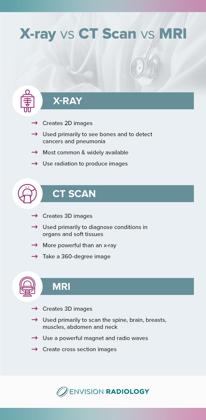
CT provides 3D cross-sections compared to the 2D flat images of traditional X-rays. CT offers high detail with layered views, while X-ray is limited with overlapping structures. Radiation exposure is moderate to high for CT and low for X-ray. CT is best for trauma, tumors, and internal organs, while X-ray is best for broken bones and chest screening. CT eliminates the superimposition problem of X-rays, revealing hidden injuries.
CT vs. MRI
CT uses X-rays while MRI uses magnetic fields and radio waves. CT involves radiation while MRI involves none. CT scans take seconds to minutes, while MRI takes 15 to 60 minutes. MRI offers excellent soft tissue detail while CT offers moderate detail. CT provides superior bone imaging while MRI provides poor bone imaging. CT has low claustrophobia risk with an open bore, while MRI has high risk with a narrow tunnel. CT is safe with metal implants, while MRI is unsafe with pacemakers. CT wins in speed and accessibility, while MRI offers better soft tissue contrast.
When CT Is the Preferred Choice
CT is preferred when speed is critical, such as in stroke or trauma situations. It is also preferred when bone detail is needed for fractures or spine issues, when patients have metal implants, or when MRI is unavailable or contraindicated.
What Patients Experience During a CT Scan
During the Scan
You will lie still on the table as it slides into the scanner. You will hear whirring or buzzing as the gantry rotates. Communication is maintained via intercom, and staff watch from a shielded control room. Children or anxious patients may receive sedation or have a parent nearby wearing a lead apron.
How Long Does It Take
A non-contrast scan takes 5 to 10 minutes. A contrast-enhanced scan takes 10 to 30 minutes. A whole-body trauma scan takes under 5 minutes. Breath-holding instructions help reduce motion blur.
Understanding Radiation Exposure
CT involves ionizing radiation, but the risk is low compared to the diagnostic benefit. Average effective doses include approximately 2 mSv for a head scan, 7 mSv for a chest scan, 10 mSv for an abdomen/pelvis scan, and 12 to 20 mSv for a whole-body scan. This compares to natural background radiation of approximately 3 mSv per year. The ALARA principle, As Low As Reasonably Achievable, guides all CT use. The estimated lifetime cancer risk from one CT scan is less than 1 in 2,000, far outweighed by the benefits when medically justified.
Modern Technological Advances in CT
Multi-Detector CT Systems
Modern MDCT scanners with 64 slices and above offer sub-second rotations, thinner slices down to 0.5 mm, cardiac imaging without motion blur, and faster trauma scans. These improvements enhance both speed and diagnostic accuracy.
Dual-Energy CT Capabilities
By using two different X-ray energies, dual-energy CT can distinguish materials, remove bone from CT angiograms, and quantify tissue composition such as liver fat or iron. This leads to more precise diagnoses without additional scans.
Iterative Reconstruction Benefits
Replacing older filtered back projection methods, iterative reconstruction reduces image noise, allowing up to 50% lower radiation doses while maintaining clarity. This technology represents a major advancement in patient safety.
Wide-Bore Scanner Designs
Newer scanners feature larger openings and shorter tunnels, improving comfort for bariatric patients, claustrophobic individuals, and those needing specialized positioning.
Quality Control and Scanner Calibration
Phantom Testing for Accuracy
CT scanners undergo regular quality assurance using a water-filled phantom with resolution targets. This test checks HU accuracy, spatial resolution, and detector consistency. Water should read 0 HU. Deviations signal hardware issues needing repair.
Laser Alignment Verification
Lasers ensure precise patient and phantom placement. Misalignment can lead to incorrect slice locations or missed pathology. Regular calibration maintains image reproducibility, essential for tracking disease over time.
Frequently Asked Questions About Cat Scanners
What is the difference between a CAT scan and a CT scan?
CAT and CT refer to the same imaging procedure. CAT stands for Computed Axial Tomography, while CT stands for Computed Tomography. CT is the modern clinical standard term, though CAT remains common in everyday usage.
How does a cat scanner produce cross-sectional images?
A cat scanner rotates an X-ray source around the body while detectors capture radiation that passes through tissues. The computer then uses reconstruction algorithms to calculate tissue density at thousands of points, creating detailed slice-by-slice images rather than overlapping 2D projections.
Is a CT scan safe?
CT involves ionizing radiation, but the doses are low enough that benefits typically outweigh risks. The estimated lifetime cancer risk from a single CT scan is less than 1 in 2,000. Modern dose-reduction techniques, including iterative reconstruction and automatic exposure control, minimize radiation exposure.
What do I need to do to prepare for a CT scan?
Preparation depends on whether contrast is used. For contrast scans, you will typically fast for 4 to 6 hours. Remove all metal objects and wear a hospital gown. Inform the technologist of any allergies, especially to iodine or shellfish, and mention any kidney problems or medications you are taking.
How long does a CT scan take?
A non-contrast CT scan takes 5 to 10 minutes. A contrast-enhanced scan takes 10 to 30 minutes. The actual scanning portion often takes only seconds to a few minutes, with most of the time spent on preparation and positioning.
Can pregnant women have CT scans?
CT scans are generally avoided during pregnancy unless absolutely necessary due to fetal radiation exposure. If a CT scan is required, the technique is optimized and the abdomen is shielded to minimize dose. MRI is often preferred for pregnant patients when appropriate.
Key Takeaways for Understanding Cat Scanners
A CAT CT scanner works by combining rotating X-rays, high-speed detectors, and powerful computers to generate detailed cross-sectional images of the body. The process begins with an X-ray tube rotating around the patient while detectors measure radiation absorption at different tissue densities. These measurements are converted to Hounsfield Units, creating a quantitative scale that distinguishes bone, soft tissue, fat, and air. Advanced technologies like slip rings enable helical scanning, while iterative reconstruction algorithms reduce noise and radiation dose.
From detecting internal injuries in emergencies to guiding cancer treatment, CT technology has revolutionized modern medicine. Its speed, precision, and versatility make it indispensable across oncology, neurology, cardiology, and trauma care. While it involves radiation, strict safety protocols and continuous technological advances continue to reduce risks while enhancing diagnostic power. Understanding how a cat scanner works empowers patients and professionals alike to make informed decisions about medical care.